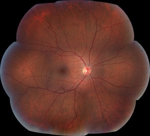
Research in the Department of Ophthalmology and Visual Sciences extends beyond the laboratory to a clinical research lab, the Fundus Photograph Reading Center (FPRC). The FPRC isa retinal imaging lab established by Dr. Matthew Davis in 1970 in order to independently analyze – or “read” – photographs from participants in the first clinical trials of diabetic retinopathy. Today, the FPRC continues to analyze retinal images providing an unbiased and comprehensive source of data in clinical trials of diabetic retinopathy, diabetic macular edema, macular degeneration, retinal vein occlusion, uveitis, inherited retinal diseases and drug safety trials.The FPRC provides this top quality retinal imaging data to researchers worldwide. “Our aim is to provide an independent source of imaging data – we evaluate retinal images for trials with small numbers of patients as well as trials with thousands of patients. In addition, we collaborate with the study leaders to analyze the results,” said Barbara Blodi, MD, professor, retina specialist and medical director of the FPRC.

“Based on our wide range of clients including the National Institutes of Health, pharmaceutical companies, small bio-tech startups and individual researchers, we know our work is making a difference on many different levels.”
From the FPRC’s inception, staff has collaborated with clinical researchers from the NationalEye Institute to create disease-specific severity scales. Importantly, these scales are used to determine both the severity of a disease and the individual patient’s prognosis. This work began with Dr. Matthew Davis who developed the diabetic retinopathy severity scale in the 1970s.This scale is based solely on retinal photographs and remains the gold standard for the ocular assessment of patients with diabetes.
The FPRC staff consists of a dedicated academic team of both certified readers and photographers all of whom have longstanding expertise in evaluating retinal images and imaging systems. FPRC Research Director, Amitha Domalpally MD, explains. “As we interpret each retinal image, our goal is to identify any changes from the normal retinal structure. FPRC staff use well-developed grading protocols and disease classifications in evaluating each individual image.” In addition to the reading of images, the FPRC provides sponsors with guidance on what imaging tests would be most beneficial and how to interpret the imaging data. “Our scientific input provides a lot of clarity to our clients,” Domalpally said. The FPRC research team is backed up by administrative staff within the Department of Ophthalmology and Visual Sciences.
Through its research, the FPRC supports the academic mission of the department and the university for students photo essay. Both Blodi and Domalpally, along with retina faculty Michael Altaweel, MD, and Mihai Mititelu, MD, MPH, serve as the principal investigators for the imaging studies performed at the Reading Center. As part of its academic mission, the FPRC routinely involves medical students, residents and fellows in the development of new measurement tools. Innovation at the FPRC is currently focused on accurately identifying and measuring retinal features on new retinal cameras and retinal scans – specifically wide-field retinal imaging and optical coherence tomography angiography. These new retinal imaging techniques require advanced measurement tools in order to provide more information on the structure and function of the retina – this, in turn, helps sponsors determine whether or not a treatment is beneficial.
Over the past 50 years the FPRC has made major contributions to ophthalmic clinical trials and has produced landmark changes in the treatment of all-too-common diseases such as diabetic retinopathy, macular degeneration, retinal vein occlusion and uveitis. However, Blodi notes that “our work is not yet done as many patients worldwide are still suffering vision loss from retinal disease.”With that in mind, Blodi and the FPRC staff will continue the momentum of the first five decades in fulfilling Dr. Davis’ original vision to foster retinal research and to support clinical investigators around the world.
Hvis vi har brug for at købe et stort antal lægemidler detaljer af vitale årsager, kan vi organisere købet gennem en medicinsk institution eller en fond. Dette er muligt for både registrerede og ikke-registrerede lægemidler.