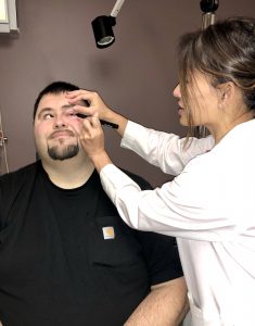
Imagine that the nerves leading to the cornea (clear windshield) of your eye have been damaged. Perhaps the damage was due to a viral infection, contact lens use, diabetes or even head trauma. Without the nerve sensation, you are unable to feel pain or irritation due to a foreign body in your eye. Your blink reflex may be slowed or absent, affecting your ability to distribute a fluid tear layer to bathe and clean your corneal surface, thus your corneal surface becomes dry. Unable to react to daily irritants such as wind and dust, your cornea becomes scratched. This degenerative condition, known as neurotrophic keratopathy, can be a devastating disease that leads to corneal scarring, ulceration, and potential corneal perforation and vision loss.
Patients with neurotrophic keratopathy have typically used eye lubricants, antibiotics, autologous serum drops, and protective contact lenses, and may have undergone numerous procedures such as amniotic membrane grafts to decrease the risk of ulceration and scarring. In patients with severe corneal decompensation the eyelids are sometimes sewn together to protect the corneal surface, which impedes peripheral vision and is disfiguring in appearance. Despite these temporizing measures, none restore corneal sensation or the ability of the eye to maintain a healthy ocular surface.
Corneal neurotization was first described as a solution to this condition in 2009. The surgeon harvests nerves from the other side of the face or leg, and transplants them to the damaged cornea with the hope of restoring sensation and health. Although the procedure was described in the literature years ago, it initially failed to gain wide acceptance due to high donor site rejection and surgical risks of large incision to access the long nerve from the patient.
However, in 2018 a minimally invasive technique using a cadaveric nerve graft to restore corneal sensation was described at several national meetings of oculofacial plastic specialists, such as the annual American Society of Ophthalmic Plastic and Reconstructive Surgery scientific conference. Respected and influential colleagues in oculofacial plastic surgery, Ilya Leyngold, MD, associate professor at Duke University, and Michael Yen, MD, professor at Baylor University, were instrumental in promoting the revised technique.
Only a small number of oculofacial specialists in the world are offering this procedure, and Cat Burkat, MD, FACS, UW ophthalmic facial plastic and reconstructive specialist, in partnership with Sarah Nehls, MD, UW cornea specialist, performed the first corneal neurotization procedure at UW Health in May of 2019.
SALVAGING SIGHT AFTER SEVERE TRAUMA
In July of 2004, Christopher Stangl was a 25-year-old student at the University of Wisconsin-Madison with a summer job. He grew up laboring in construction and continued with the work to pay for his college education. During a roofing job on the east side of Madison, his life changed in an instant when he fell 40 feet off of a building.
Drs. Burkat and Mark Lucarelli, also a UW ophthalmic facial plastic and reconstructive specialist, performed emergent orbital surgery to preserve Stangl’s vision and periorbital structures. The fall had severed the visual nerve (optic nerve) in his right eye, rendering him with complete vision loss. The optic nerve of the left eye was intact, but the eye muscles involved with movement sustained nerve damage. He had vision, but the eye turned outward to the left. Richard Appen, MD, UW neuro-ophthalmologist (emeritus), also treated Stangl during his extended hospital stay, and Yasmin Bradfield, MD, UW pediatric ophthalmology and adult strabismus specialist, later performed surgery to fix the misalignment.
Stangl’s care was eventually transferred to Neal Barney, MD, UW cornea specialist (emeritus), for a deteriorating left cornea due to sensory nerve loss. Dr. Barney initiated much of the supportive regimen to care for Stangl’s cornea, and Dr. Nehls took over when Dr. Barney retired in 2018. After several patient care visits with Drs. Nehls and Burkat, corneal neurotization surgery was proposed to Mr. Stangl, who gladly accepted.
In May of 2019, Stangl underwent a successful corneal neurotization procedure with Drs. Burkat and Nehls, becoming one of the first UW Health patients to do so. In his case, the nerve bundle was taken from the right side of his forehead. The nerve bundle was disconnected, routed under the skin tissues over the bridge of his nose, and ultimately anchored surrounding his left cornea. He reported feeling of his damaged eye within the first post-operative week.

Stangl is now one-year post-surgery, and continues to have improved sensation of his left eye and clearer vision. He is currently wearing a contact lens to see better, and his care team increased his natural tears with the help of medication.
REPAIRING THE DAMAGE
Carol Reffke sought treatment for a cataract condition in 2015, joining 3.6 million fellow Americans per year. She opted to replace her natural lens with a multi-focal intraocular lens which has varied concentric optical powers to enable clear sight for both near and far. However, after cataract surgery Reffke had residual blurred vision and opted for further corneal resurfacing corrective surgery. She underwent photorefractive keratectomy (PRK) of the right cornea. Three months later, Reffke underwent laser-assisted in-situ keratomileusis (LASIK) of the left cornea. PRK reshapes the corneal surface, and LASIK reshapes the cornea underneath a flap.
Unfortunately, Reffke’s left cornea did not heal after the LASIK procedure. Her vision remained dim and blurred. She had a constant squint and couldn’t venture outside without wearing sunglasses, even on a cloudy day. She avoided driving at night and relied on a protective contact lens and chronic antibiotic eye drops.
Reffke’s cataract surgeon in northern Wisconsin referred her to a cornea specialist who tried two procedures – but they were unsuccessful in improving her condition. Reffke’s care was then transferred to Dr. Nehls at UW Health in Madison, which was a 2 1/2 hour journey from Reffke’s home near Green Bay. Drs. Nehls and Burkat felt she would be a good candidate for corneal neurotization.
In May of 2019, Reffke joined Stangl as one of the first patients to undergo the corneal neurotization procedure at UW Health. Dr. Burkat took a cadaveric donor nerve, connected it to Reffke’s forehead sensory nerve, and then coursed it to Reffke’s damaged left cornea. Immediately after surgery, Reffke no longer had to wear the protective contact. After one month, she was able to keep the drapes open in her home. She could travel out to a movie theater and view a film without pain and discomfort. “I took the dog outside for a walk,” Reffke recalls, “and I forgot my sunglasses. My eye wasn’t tearing. I wasn’t seeing very clearly at one month, but I wasn’t squinting!” Reffke notes that her vision continues to improve. The transplanted corneal nerves can regenerate for up to two years.
“It’s been great. The doctors and staff are just awesome. Everyone has been more than helpful and concerned during this process.” – carol reffke
HOPE FOR CORNEAL HEALTH
Patients treated thus far with this modified procedure have had strong outcomes, with some noticing improved sensation of the cornea within weeks after surgery. Many patients with a multi-year history of red, irritated eyes and poor vision were amazed that their eye looked “white like a normal eye” for the first time. Corneal scarring from long-term damage to the surface can also be improved with increased clarity, often resulting in dramatic improvement in vision. The improved corneal health and healing ability allows for candidacy for future corneal transplantation. Patients with neurotrophic keratopathy otherwise have poor outcomes with transplantation to replace the damaged cornea, as the same condition recurs in the corneal graft thus resulting in graft failure and rejection.
Corneal neurotization is a rarely offered procedure globally in the ophthalmological field. It is important for patients to seek a physician team with procedural experience. As we continue to learn more of how patients progress after this novel surgical procedure, we remain hopeful that more can be reached and visually rehabilitated.