a New Way of Imaging
One of the most exciting new projects in vision research at UW-Madison is a groundbreaking group collaboration among the McPherson Eye Research Institute, the Department of Ophthalmology and Visual Sciences, and other UW and national partners. The Wisconsin Advanced Imaging of Visual Systems (WAIVS) Lab, founded in 2018, promises to establish and advance exciting new eye imaging techniques at UW-Madison.
Page through any science magazine, and you’ll be amazed at the image quality that is attainable by today’s equipment – whether of distant galaxies or within the human body. Imaging techniques developed in recent decades and used to image the eye and retina, such as Optical Coherence Tomography (OCT), have advanced both patient care and basic research immensely. It is a paradox, though, that the more we see, the more we realize how much more there is to see. For instance, we now have the ability to view images of living photoreceptor cells with better clarity than ever before; however, we are only at the starting line when it comes to peering within those cells and understanding the functional relationships of the various cell components. Understanding those relationships is vital to fighting disease and devising better therapies.
![]()
The WAIVS project brings together a group of outstanding engineers, basic science researchers, and clinicians who are building an imaging lab featuring an Adaptive Optics (AO) platform. Adaptive Optics is a recently-developed method of eliminating aberrations in images by using dynamic mirrors that correct distortion. The same technology has been used widely in astronomy for improving images from telescopes. WAIVS will have two Adaptive Optics Scanning Laser Ophthalmoscope (AOSLO) machines housed in dedicated McPherson ERI space in the Wisconsin Institutes for Medical Research (WIMR). One machine will be used for patient research, allowing researchers and clinicians to understand retinal dysfunction on a cellular level in living patients, and also to monitor cellular responses to treatments such as stem cell and gene therapies. In a separate, but nearby space, the other AOSLO machine will be used for building and testing innovative new features for the existing technology that will allow us to “see further.” Having the two machines in close proximity will allow for rapid application of imaging breakthroughs.
What can we see with Adaptive Optics?
Optical coherence tomography (OCT), as shown in images A-C, has become an invaluable tool for clinical imaging of healthy or diseased retina. OCT provides 3D volumetric images that can then be inspected from different angles. At top, panels B and C are cross-sections corresponding to the dashed lines in panel A. Despite the clear value of OCT imaging, resolution of OCT remains limited due to imperfections in the lens, cornea, and tear film of the subject. Adaptive Optics solves this limitation, providing “fine focus” with cellular resolution as shown in panel D that corresponds to the tiny square in panel A. Even with this resolution, cells can be difficult to visualize using standard confocal contrast. State of the art AOSLO systems like the one being developed at UW-Madison include additional split-detector capabilities as shown in panel E to improve contrast and visualization of photoreceptors. In this image, it is possible to see the abnormal morphology of most of the visible photoreceptor cells.
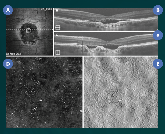
Image adapted from “Photoreceptor Inner Segment Morphology in Best Vitelliform Macular Dystrophy,” Retina. 2017 Apr; 37(4): 741–748.
To better understand WAIVS, we’d like you to meet some of its team members.
Jeremy Rogers, MS, PhD, (Biomedical Engineering) is the principal UW-Madison investigator responsible for the fabrication and innovation of the AOSLO machines, which are being housed within his lab space in WIMR. He will also oversee advancements in parallel technologies that need to grow with improvements in AOSLO capabilities. With experience in multiple advanced imaging techniques, Dr. Rogers aims to advance AOSLO and other eye and retinal imaging systems to make them as effective as possible.
Alfredo Dubra, PhD, (Ophthalmology, Standford University) and Joseph Carroll, PhD, (Ophthalmology, Medical College of Wisconsin) have been at the forefront of building and applying AOSLO technology from its inception, and will collaborate closely in its installation and use at UW-Madison. These AOSLO systems are extraordinarily delicate and will be constructed on vibration isolation optical tables using a combination of custom-made and off-the-shelf equipment. Drs. Dubra and Carroll will train the UW-Madison team and serve integral roles on the WAIVS team.
Kimberly Stepien, MD, (DOVS) will direct the use of AOSLO equipment in investigating human ocular diseases, including macular degeneration and inherited retinal conditions such as retinitis pigmentosa. Of great importance is the development of techniques to monitor the effectiveness of new therapeutic approaches, such as gene and stem cell-based therapies, that will be developed and tested at UW-Madison in coming years.
Barbara Blodi, MD, (DOVS) will oversee the interface between the WAIVS Lab and the Fundus Photograph Reading Center (FPRC), a unit of the Department of Ophthalmology and Visual Sciences that is world-renowned for its expertise in retinal imaging analysis. The FPRC will help develop standardized Adaptive Optics imaging and grading protocols for image analysis. This is necessary in order for data to be correlated across AO research projects and with other imaging methods.
David Gamm, MD, PhD, (DOVS) will focus on the potential use(s) of AOSLO imaging in clinical trials for stem cell-based therapies to treat age-related macular degeneration and retinitis pigmentosa.
These researchers are a subset of those working to build the WAIVS lab; others include Kevin Eliceiri, PhD, (Medical Physics; Morgridge Institute for Research; associate director of the McPherson Eye Research Institute), Gillian McLellan, BVMS, PhD, (DOVS; School of Veterinary Medicine) and Melissa Skala, PhD, (Biomedical Engineering; Morgridge Institute for Research). It is truly a cross-campus team whose common goal is to bring state-of-the-art ophthalmic imaging to UW-Madison and combine it with our formidable know-how to make it even more informative and – ultimately – beneficial to patients.
Original Source: McPherson Eye Research Institute